
| name | Amanita peltigera |
| name status | nomen acceptum |
| author | D. A. Reid |
| english name | "Shield-Bearing Death Cap" |
| images |
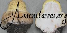
 1. Amanita peltigera, Wungong Catchment, West Australia, Australia  2. Amanita peltigera, Wungong Catchment, West Australia, Australia |
| intro | The following description is based on Reid (1980). The description of this species is based on the only known specimen. |
| cap | The cap of Amanita peltigera is 55 mm wide, plano-convex, with a smooth and naked margin. The gray cap is almost entirely covered by a very large, single, roughly circular, white patch of volval tissue. |
| gills | No information recorded. |
| stem | The stem is 50 × 10 mm, with a swollen rooting base, white with a hint of grayish penciling. No annulus was found on the type specimen. It is not clear from the original description whether the volva is truly saccate or only limbate. The presence of a large bulb suggests the latter is the case, and this is confirmed by my examination of the type at Kew. |
| odor/taste | No information recorded. |
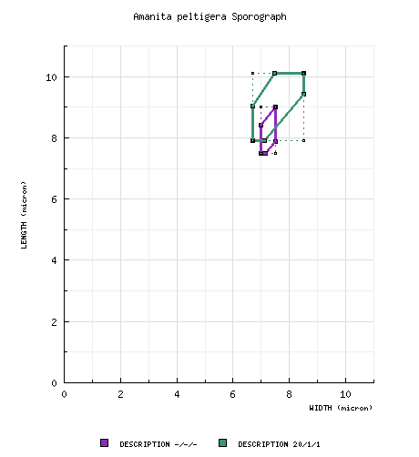
| spores | According to Reid, the spores of this species measure (6.5-) 7.5 - 9.0 x (6.0-) 7.0 - 7.5 (-8.5) µm and are subglobose or ovate to very broadly ellipsoid (infrequently globose, infrequently ellipsoid) and amyloid. Clamps are absent at base of basidia. My measurements of spores from the type specimen are (7.6-) 7.9 - 10.1 (-11.9) × (6.3-) 6.7 - 8.5 (-9.1) µm. |
| discussion |
This species is originally described from the state of Western Australia. No ecological information was recorded. Reid does not discuss placement of this species in a section. Wood (1997) proposes section Validae. The absence of an annulus is problematic for Wood's choice. Reid's illustration showing thick-walled hyphae dominating the volva (I found thick-walled elements rather common in the volva, but not to the extent of Reid's illustration) reminds one of the similar hyphae in several species of section Phalloideae. My examination of the type shows that the volva is not as extensively layered as are the volvas in sect. Amidella. A membranous volva is unknown in section Validae. If we take into account recent molecular work supporting Bas' (1969) hypothesis that section Phalloideae may have arisen from an ancestor that could be placed in section Amidella, then the possibility must be acknowledged that a mushroom assignable to section Phalloideae might exist that lacks a well-formed annulus. It is also possible that there was an annulus that was lost from the type specimen of A. peltigera.—R. E. Tulloss |
| brief editors | RET |
| name | Amanita peltigera | ||||||||
| author | D. A. Reid. 1978. Victorian Naturalist 95: 49. | ||||||||
| name status | nomen acceptum | ||||||||
| english name | "Shield-Bearing Death Cap" | ||||||||
| etymology | peltus, "shield" + -ger, "-bearing" (adjectival ending); hence, "shield bearing," because of the large patch on the cap of the holotype. | ||||||||
| MycoBank nos. | 308577 | ||||||||
| GenBank nos. |
Due to delays in data processing at GenBank, some accession numbers may lead to unreleased (pending) pages.
These pages will eventually be made live, so try again later.
| ||||||||
| holotypes | K | ||||||||
| type studies | Tulloss (partial) herein | ||||||||
| revisions | Reid. 1980. Austral. J. Bot., Suppl. Ser. 8: 47, figs. 31(a-b), 87. | ||||||||
| intro |
The following text may make multiple use of each data field. The field may contain magenta text presenting data from a type study and/or revision of other original material cited in the protolog of the present taxon. Macroscopic descriptions in magenta are a combination of data from the protolog and additional observations made on the exiccata during revision of the cited original material. The same field may also contain black text, which is data from a revision of the present taxon (including non-type material and/or material not cited in the protolog). Paragraphs of black text will be labeled if further subdivision of this text is appropriate. Olive text indicates a specimen that has not been thoroughly examined (for example, for microscopic details) and marks other places in the text where data is missing or uncertain. The following material is derived from the protolog of the present taxon and from original research by R. E. Tulloss. Because the partial type study of Tulloss was carried out in 1988 prior to his differentiation of vascular hyphae from other hyphae that appear refractive, the editors choose to use the more general term "refractive hyphae" in reporting his results. | ||||||||
| pileus | from protolog: 55 mm wide, gray, plano-convex; context not decribed; margin "smooth and naked"; universal veil as single, very large, white, patch, with surface cracking into large separable membranous placques "seated on a continuous felt-like base." | ||||||||
| lamellae | not described in protolog. | ||||||||
| stipe | from protolog: 50 × 10 mm, white, with "hint of greyish pencilling"; bulb "swollen rooting," up to to 23 mm wide; context not described; partial veil lacking; universal veil "sheathing," whitish, "saccate." | ||||||||
| odor/taste | not recorded. | ||||||||
| macrochemical tests |
none recorded. | ||||||||
| pileipellis |
not described in protolog. from type study of RET: filamentous undifferentiated hyphae 6.2 - 7.3 μm wide, dominant, branching, lacking subradial arrangement; refractive hyphae 3.6 - 8.5 μm wide, branching, rather common. | ||||||||
| pileus context | not described in protolog. | ||||||||
| lamella trama |
not described in protolog. from study of type by RET: bilateral, divergent; ??; ??; refractive hyphae 2.4 - 5.1 μm wide, branching, rather plentiful. | ||||||||
| subhymenium |
from protolog: conspicuously pseudoparenchymatous from study of type by RET: pseudoparenchymatous (cellular); comprising globose to subglobose to ellipsoid inflated cells, ca. 40 μm deep, with some basidioles extending about 20 μm into subhymenium | ||||||||
| basidia |
from protolog: 30 - 37 × 9 - 10 μm; clamps lacking. from study of type by RET: 38 - 55 × 10.9 - 15.8 μm, dominantly 4-, also 2- and 3-sterigmate; clamps not observed. | ||||||||
| universal veil |
from protolog: hyphae 3 - 12 μm wide, predominating, hyaline, branched, with thin but distinct walls, often somewhat inflated; inflated cells extremely scanty, subglobose or ovoid, up to 110 × 90 μm; clamps lacking. from type study of RET: On stipe base, exterior surface: filamentous undifferentiated hyphae 2.6 - 9.7 μm wide, dense, often longitudinally oriented, dominant; inflated cells (even very near surface), subclavate to ventricose, thin-walled, frequent, up to 57 × 27 μm; refractive hyphae 7.3 - 8.5 μm wide. On stipe base, interior: filamentous undifferentiated hyphae 5.7 - 15.8 μm wide, densely interwoven, thin-walled, curving but not coiling; inflated cells same shapes as at surface, thin-walled, up to 99 × 35 μm, also subglobose (e.g., 27 × 26 μm); refractive hyphae not observered. On pileus, within "placque": Very similar to that on stipe base perhaps with more and larger inflated cells, but still dominated by filamentous undifferentiated hyphae, uniform throughout except for exterior surface; inflated cells up to 124 × 32 μm; refractive hyphae present throughout, 3.5 - 8.5 μm wide. On pileus, from crumbly layer beyond edge of "placque": with hyphae and inflated cells about equally frequent, with many cell walls apparently thickened; tissue as elsewhere (above); filamentous undifferentiated hyphae with walls thin or (apparently) up to 0.7 μm thick; inflated cells occasionally ovoid in addition to previously reported shapes, up to 82 × 75 μm, with walls thin or (apparently) 0.5 - 1.2 μm thick. | ||||||||
| stipe context | not described in protolog. | ||||||||
| partial veil | absent. | ||||||||
| lamella edge tissue | not described in protolog. | ||||||||
| basidiospores |
from protolog: [-/-/-] (6.5-) 7.5 - 9.0 × (6.0-) 7.0 - 7.5 (-8.5) μm, (est. Q = 1.05 - 1.20), amyloid, subglobose to broadly ellipsoid; apiculus not recorded; contents not recorded; color in deposit not recorded. from type study of RET: [20/1/1] (7.6-) 7.9 - 10.1 (-11.9) × (6.3-) 6.7 - 8.5 (-9.1) μm, (L = 9.2 μm; W = 7.5 μm; Q = (1.02-) 1.11 - 1.35 (-1.38); Q = 1.23), ??, smooth, thin-walled, amyloid, subglobose to broadly ellipsoid to ellipsoid, infrequently globose, somewhat adaxially flattened; apiculus sublateral, truncate conic, proportionately small; contents ??; color in deposit unknown. | ||||||||
| ecology | not described in protolog. | ||||||||
| material examined | from protolog and type study of RET: AUSTRALIA: WESTERN AUSTRALIA—Stirling West, iii.1976 John Randals s.n. (holotype, K). | ||||||||
| discussion |
Because of the absence of a partial veil in the single specimen of the type, there may be some hesitation to place this species in Amanita sect. Phalloideae; but, at present, that seems to be the best choice considering the microscopic structure that has been reported for the universal veil. A distinct bulb at the stipe base and the lack of multiple, distinct layers in the interior of the universal veil appear to eliminate inclusion of the present species in sect. Amidella. The word "apparent" is used in reference to thickness of cell walls because collapsed cells can appear to have thickened walls when uncollapsed cells are clearly thin-walled. This fact may not have been clear to RET in 1988. On the other hand, Reid's fig. 87 (1980) shows thickened walls on hyphae in an unidentified location within the universal veil even though the 1980 text seems to imply that he didn't see thickened walls in that tissue. | ||||||||
| citations | —R. E. Tulloss | ||||||||
| editors | RET | ||||||||
Information to support the viewer in reading the content of "technical" tabs can be found here.
Each spore data set is intended to comprise a set of measurements from a single specimen made by a single observer; and explanations prepared for this site talk about specimen-observer pairs associated with each data set. Combining more data into a single data set is non-optimal because it obscures observer differences (which may be valuable for instructional purposes, for example) and may obscure instances in which a single collection inadvertently contains a mixture of taxa.

Text and User-Generated Sporographs are published under the Creative Commons License.

In the case of a taxon page, image credits are on the 'image' tab.